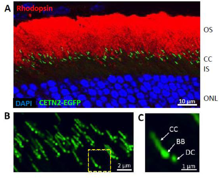
Figure 7.
Transgenic expression of EGFP-CETN2 identifies photoreceptor centrioles and connecting cilia. A, rod outer segments labeled with anti-rhodopsin (red), centrioles expressing EGFP-CETN2 (green) and nuclei binding DAPI (blue). B, confocal microscopy of several connecting cilia. C, a single photoreceptor connecting cilium streak resembles the tail of a shooting star. BB, basal body; DC, daughter centriole.

