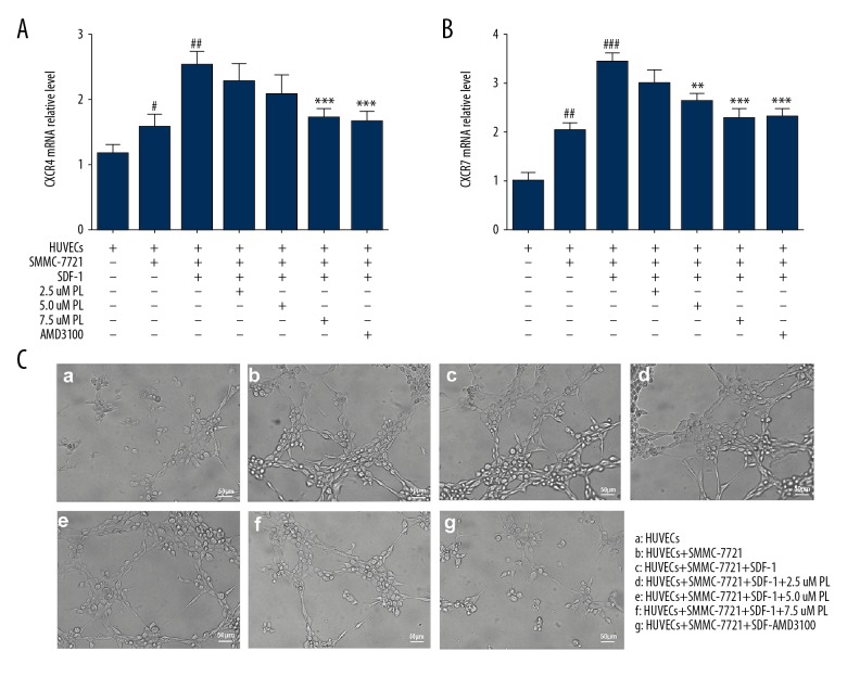
Figure 3.
Effect of PL on the mRNA expression of CXCR4 and CXCR7 genes in SMMC-7721 cells co-cultured with HUVECs, grouped as follows: HUVECs only; HUVECs were co-cultured with SMMC-7721 cells; HUVECs were co-cultured with SMMC-7721 induced by SDF-1; the SMMC-7721 cells were treated with 2.5–7.5 μM PL as indicated; AMD3100 (100 ng/ml) was used as the positive control. After being exposed to the indicated concentrations of PL and AMD3100 for 24 h, the mRNA expression of CXCR4 and CXCR7 were analyzed using quantitative real-time PCR. Each bar represents the mean value ± standard deviation (SD). * P<0.05; ** P<0.01; *** P<0.001, compared to the HUVEC group. # P<0.05; ## P<0.01; ### P<0.001, compared to the SDF-1 group (A, B). Microscopically co-cultured HUVECs spontaneously form capillary-like structures on Matrigel, particularly SDF-1 induced SMMC-7721 cells. The number and continuity of capillary-like structures in HUVECs were significantly inhibited by 2.5–7.5 μM PL in a dose-dependent manner (scale bar, 50 μm) (C).

