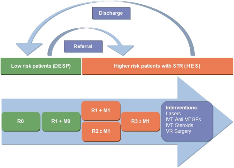
Fig. 1.
Patient flow diagrama. aSee Table A1 for more details on classification of diabetic retinopathy. R0, no retinopathy; R1, background retinopathy; R2, pre-proliferative retinopathy; R3, proliferative retinopathy; M0, no maculopathy; M1, maculopathy present; DESP, diabetic eye screening programme; HES, hospital eye services; STR, sight-threatening retinopathy; IVT, intra-vitreal therapy; VEGF, vascular endothelial growth factor; VR, vitreoretinal

