Fig. 5.
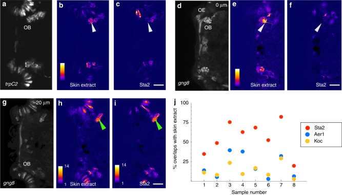
Effect of bacterial lysate on the olfactory system. a–i Calcium imaging of the olfactory bulb two different larval fish exposed to skin extract (b, e, h) or bacterial lysate (c, f, i). Average projection of the time-series, showing GCaMP6s expression in olfactory sensory neurons and in terminals, driven by the trpc2 (a) or gng8 (d, g) promoter. Panels d–i are the same fish, with g–i showing a plane that is 20 µm deeper than d–f. A glomerulus in the dorsal bulb (white arrowhead) is activated by skin extract but not by the bacterial lysate. Both lysates activate identical terminals in the ventral bulb (green arrowhead). j Percentage of overlap between response to three different bacterial lysates and skin extract. The highest overlap is seen with lysate from Staphylococcus sp. ZWU0021. Anterior is to the left; OB olfactory bulb, OE olfactory epithelium. Scale bar = 25 µm

