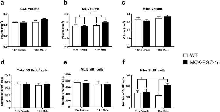
Figure 2.
Overexpression of PGC-1α in skeletal muscle does not influence hippocampal volume or cytogenesis in middle-aged animals. Graphs showing volumes of DG subregions (a) GCL (two-way ANOVA; n = 8–10; n.s.), (b) ML (two-way ANOVA, n = 8–10; sex effect, *p < 0.05; genotype and interaction effect, n.s.), and (c) hilus (two-way ANOVA; n = 8–10; n.s.) in 11-month-old female WT and transgenic animals. Graphs showing corresponding number of BrdU+ cells in the (d) DG, (e) ML and (f) hilus (two-way ANOVA; n = 9–14; genotype effect, *p < 0.05) 4 weeks after the first BrdU-injection. Data expressed as mean ± standard error of the mean (SEM) for WT and MCK-PGC-1α animals.

