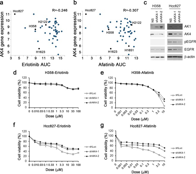
Figure 5.
AK4 signaling helped the response of EGFR inhibitors. (a) Correlation plot of AK4 expression level and AUC of Erlotinib in COSMIC lung adenocarcinoma cell lines. Specific cell lines were presented in black. (b) Correlation plot of AK4 expression level and AUC of Erlotinib in COSMIC lung adenocarcinoma cell lines. Specific cell lines were presented in black. (c) The expression level of AK1, AK4, pEGFR, and EGFR in H358 and Hcc827 cells after shAK4 treatment. Beta-actin was used as loading control. (d) The cell viability assay of Erlotinib in H358-shLuc, H358-shAK4-1 and H358-shAK4-2 cells. (e) The cell viability assay of Afatinib in H358-shLuc, H358-shAK4-1 and H358-shAK4-2 cells. (f) The cell viability assay of Erlotinib in Hcc827-shLuc, Hcc827-shAK4-1 and Hcc827-shAK4-2 cells. (e) The cell viability assay of Afatinib in Hcc827-shLuc, Hcc827-shAK4-1 and Hcc827-shAK4-2 cells.

