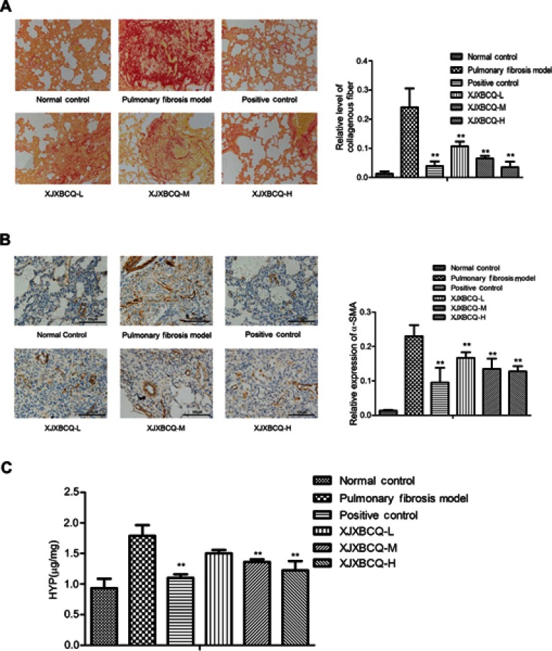
Figure 3.
Effect of XJXBCQ on PF in vivo. (A) Representative images of lung collagen deposition by picro-sirius red staining from rat treated with saline; BLM (7 μg/g), positive control and BLM+XJXBCQ on the 28th day. (n=5); scale bar, 200 μm. (B) Protein expression levels of α-SMA in lung tissues. Scale bar, 100 μm. (C) HYP content in lung tissues of BLM-induced rat. The values are shown as mean±SEM of the three experiments. **P<0.01 versus the model group.

