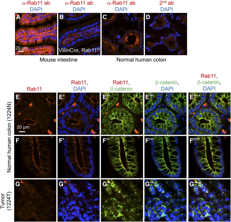
Figure 1.
Rab11 expression in mammalian intestine and colon. (A–D) Immunofluorescence staining of tissue sections using a Rab11 antibody, or secondary antibody alone in D. (A) Wild-type mouse small intestine, (B) Rab11flox/f;lox; villinCre mouse intestine, (C) normal human colon, and (D) normal human colon staining with secondary antibody alone. Red color is Rab11 staining, and blue is DAPI for DNA staining. (E–G) Immunofluorescence staining of colon tumor and matching normal tissue from a deidentified patient. The tissue sections were double stained using antibodies for Rab11 (shown in red) and β-catenin (shown in green). Blue is DAPI staining. E and F are images of different parts of the same slide, showing horizontal sections and longitudinal sections, respectively, of the colonic crypts. G is an image of a horizontal section of the tumor tissue. The different color combinations, as indicated, of the same region are shown in the parallel panels (E–E””, F–F””, and G–G””). The number 1224N and 1224T represents the sample number of the UMMS tissue bank deidentified patients, with N for normal and T for tumor.

