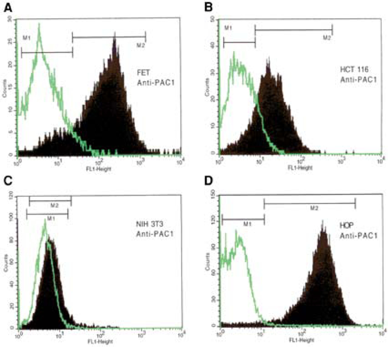
Fig. 1.

Cytofluorimetric analysis. Rabbit polyclonal anti-PAC1 antibodies followed by Alexa 488 goat anti-rabbit IgG (Molecular Probes) were used to detect the presence of PAC1 receptor on FET and HCT116 colon cancer cell lines. Panels A and B show that respectively 83.06%, and 65.28% cells were positive for PAC1 receptor expression, respectively (M2). Untransfected NIH-3T3 wild-type and PAC1-transfected NIH-3T3 cells (HOP) were used as negative and positive controls for PAC1, respectively (C,D). Rabbit IgG was used as a negative control for a specific binding (M1).
