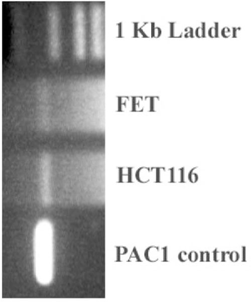
Fig. 3.

PCR analysis. PAC1 primers detected the presence of PAC1 receptor mRNAat ~1.35 kb. The primers used were (sense 1) 5′-TGCTGGCCAAGTGTCATG-3′ (nucleotides 50–70) and (antisense) 5′-CTGGGACCGCA GGTGC-3′ (nucleotides 1780–1800) (Pisegna and Wank, 1996) in FET and HCT116 cell lines. The SV1 or HIP PAC1 cDNA was used as a positive control.
