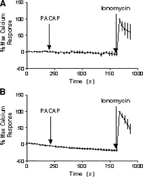
Fig. 5.

PACAP-38-induced Ca2+ to PACAP-38. FET and HCT116 cells were loaded with Fluo-4 AM for the visualization of Ca2+ transport under confocal microscopy. Atime series of a total of 40 images with 20-s intervals in between was taken to monitor the Ca2+activity. Upon activation with 2.5 × 10⁻⁷ M PACAP-38 at 200 s (tenth frame), there was no Ca2+ movement observed for both FET and HCT116 cells (A,B) relative to the response obtained from the addition of ionomycin. The addition of 10 μM ionomycin at the thirty-fifth frame of imaging induced a peak that signaled maximum calcium response.
