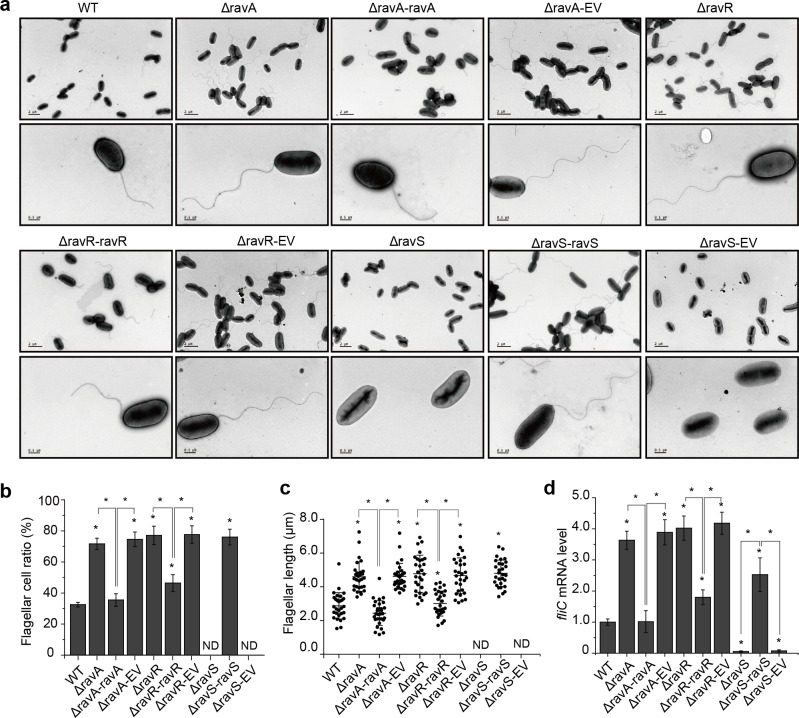
Fig 2. ravA, ravR and ravS differentially regulate the development of bacterial flagella.
(a) Morphology of bacterial flagella. Bacterial flagella were observed by transmission electron microscopy after negative staining. A representative image of each strain is shown (n > 30). Upper panel: bacterial population profile; lower panel: morphology of a single bacterium. (b) Ratio of bacteria with flagella. For each strain, cells with flagella were counted for three biological replicates with each containing at least 100 cells. (c) The average flagellar length of bacterial strains. The length was measured using the AutoCAD software. Three biological replicates were taken with each comprising at least 30 measurements. (d) The fliC mRNA level in bacterial strains. The level of fliC mRNA was measured by qRT-PCR. Amplification of cDNA from tmRNA was used as an internal control. The experiment was repeated three times and the result of a representative experiment is shown. In (b–d), standard deviations are provided; asterisk: significant difference, as tested by Student’s t-test (P ≤ 0.05).

