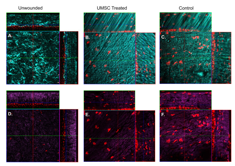
Figure 4.
Reorganization of collagen fibril architecture in UMSC-treated eyes following injury in Col5a1-deficient mice. A–C: Forward-scattered second harmonic generated (SHG) signals (cyan) depicting collagen fibrils. D–F: Backscattered SHG signals representing the overall lamellar structure of the stroma 7 days after treatment. The control eyes show enlarged collagen fibrils (C) and disorganized lamellae (F) compared to the transparent and flattened lamellae in the umbilical cord mesenchymal stem/stromal cells (UMSC)-treated eyes (B–E). Red = Syto59 nuclear stain. Seven days post-UMSC treatment. UMSC treatment more closely resembles an unwounded eye (A, D).

