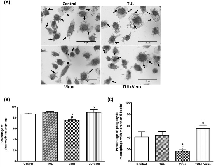
Fig 11. Tulathromycin restores Fc-mediated phagocytosis in PRRSV-infected porcine MDMs.
Opsonized phagocytosis of IgG coated latex beads in MDMs following HBSS (control) or tulathromycin (1mg/mL; TUL) treatment for 1h and infection with PRRSV (m.o.i. = 0.5; Virus and TUL+Virus) for 12 hours was measured. Following treatment and infection, MDMs were incubated with IgG coated latex beads for 45 minutes. (A) Micrographs of macrophages that have phagocytosed latex beads (arrows). Bar = 20μm (B) Percentage of macrophages that phagocytosed at least 1 latex bead. (C) Percentage of macrophages that phagocytosed at least 5 IgG-coated latex beads. n = 150–200 macrophages/group. Images and histograms are representative of 4 independent experiments. Mean ± SEM. # = P<0.05 vs Control; * = P<0.05 vs TUL; Δ = P<0.05 vs virus.

