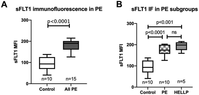
Figure 2.
Placental sFLT1 activity by immunofluorescence staining. Comparison of placental sFLT1 mean fluorescence intensity (MFI) by immunofluorescence (IF) staining in control and all PE (including both PE without laboratory abnormalities and HELLP syndrome) displayed as box plots with median with 1st and 3rd interquartile range; whiskers at the 5th and 95th percentile (A). sFLT1 MFI in control and subtypes of PE; PE without laboratory abnormalities and HELLP syndrome (B).

