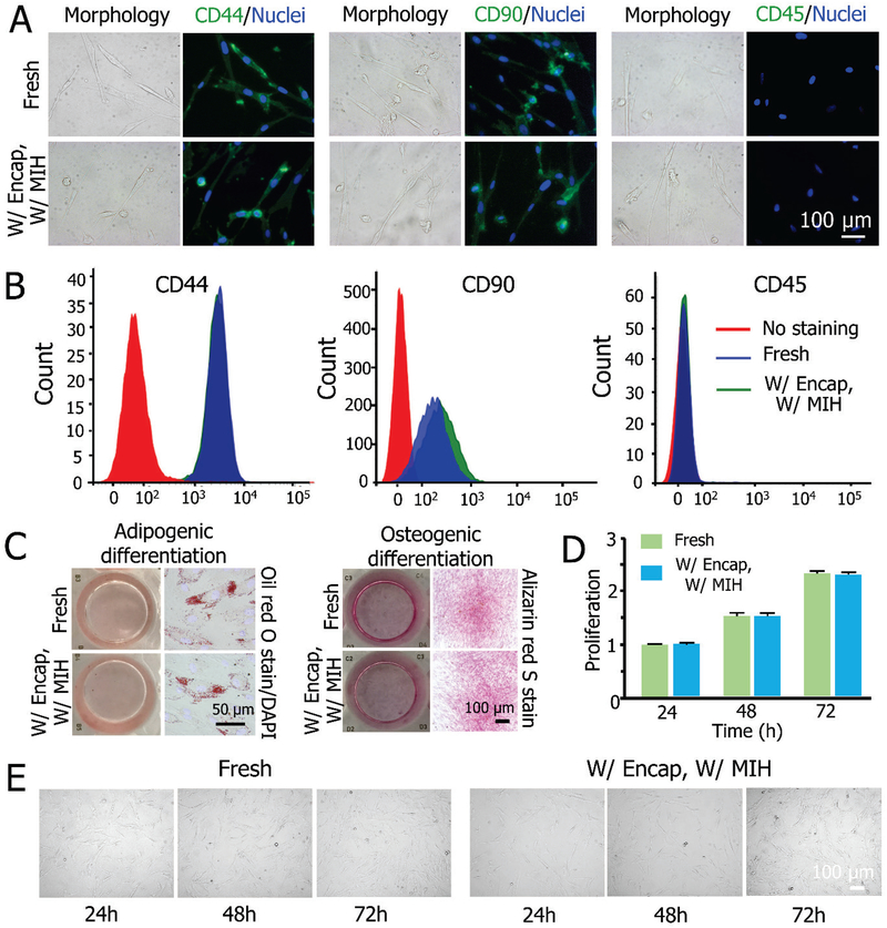
Figure 5.
Functional properties of MSCs before and after vitrification with MIH and encapsulation. A) Fluorescence immunostaining for CD44 (+), CD90 (+), and CD45 (−) showing the expression of the three markers on MSCs. B) Quantification of CD44 (+), CD90 (+), and CD45 (−) expression on the cells by flow cytometry. C) Qualitative analysis of adipogenic (left) and osteogenic (right) differentiation of MSCs post-vitrification compared to fresh cells. D) Proliferation of MSCs released from constructs post-vitrification compared to fresh cells. E) Typical DIC micrographs between cryopreserved and fresh MSCs showing the cell proliferation.

