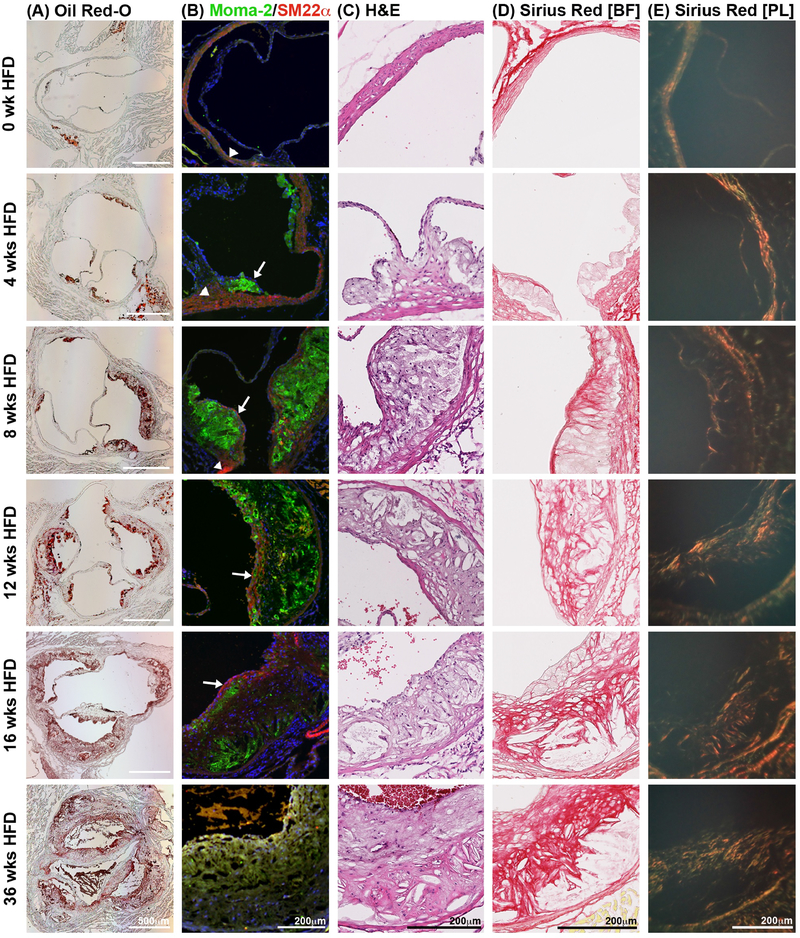
Fig. 1:
Histological analysis of the pathophysiology in the atherosclerotic plaque.
Ldlr−/− mice on HFD for several time points were euthanized and frozen aortic root sections prepared. (A) Oil Red-O staining of aortic root sections was used to determine atherosclerotic plaque size. (B) Serial sections of the aortic root were stained with the macrophage marker, Moma-2 (green), to determine macrophage content per plaque size. SM22α was used to stain smooth muscle cells (red). Nuclei shown by DAPI staining in blue. (C) H&E staining of aortic root sections allowed for the quantification of the necrotic core. (n=10 for each given time point). (D) Picro Sirius Red staining of aortic root sections allows for visualization and quantification of total collagen using bright field microscopy, and of collagen type and maturation using polarized light microscopy (E). Under polarized light, collagen III appears green while collagen I shows in yellow, orange and red in order of increasing thickness. n=3; sections from both upper and lower aortic root were analyzed; BF, bright field; PL, polarized light.

