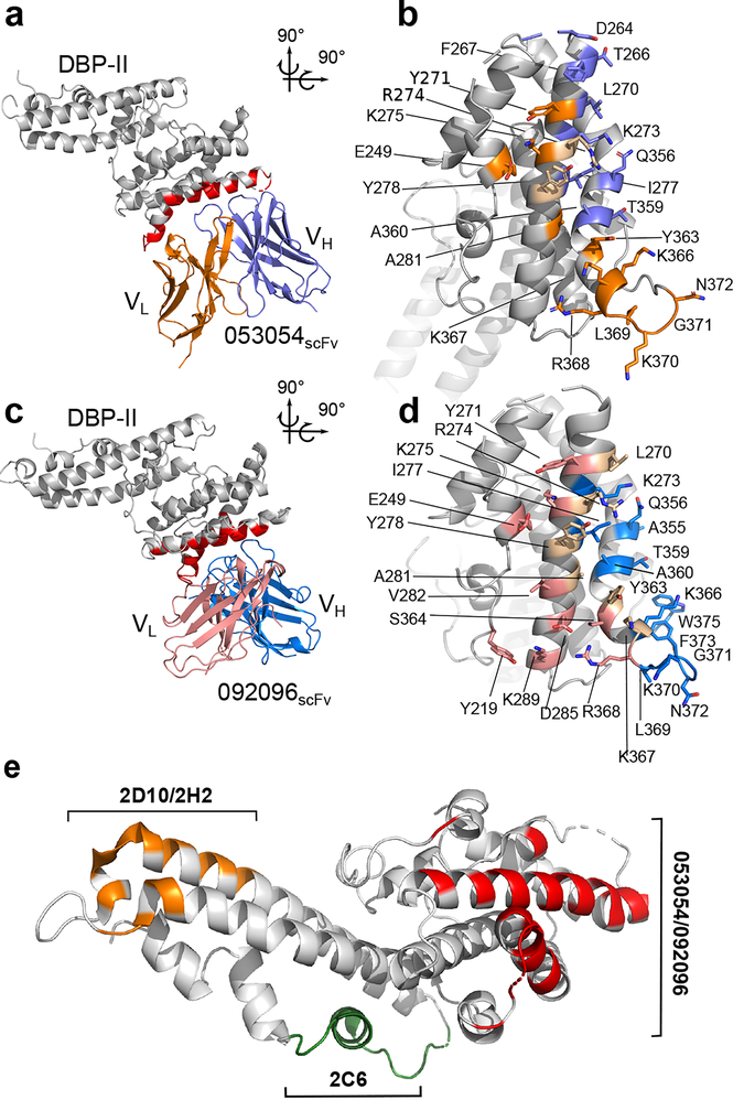
Figure 1|. Structural definition of human antibody epitopes in DBP.
a, Overall structure and epitope for the 053054 complex. Gray - DBP-II. Dark blue - 053054 heavy chain. Orange - 053054 light chain. Red - epitope. b, orthogonal detailed view of the epitope for 053054 in DBP. Gray - DBP-II. Dark blue – DBP residues contacted by the 053054 heavy chain. Orange – DBP residues contacted by the 053054 light chain. Beige – DBP residues contacted by both heavy and light chains. c, Overall structure and epitope for the 092096 complex. Gray - DBP-II. Light blue - 092096 heavy chain. Pink - 092096 light chain. Red - epitope. d, orthogonal detailed view of the epitope for 092096 in DBP. Gray - DBP-II. Light blue – DBP residues contacted by the 092096 heavy chain. Pink – DBP residues contacted by the 092096 light chain. Beige – DBP residues contacted by both heavy and light chains. e, Comparison of human and murine epitopes19 in DBP-II reveal epitopes are distinct. Orange - epitope of inhibitory murine mAbs 2D10/2H2. Green - epitope of inhibitory murine mAb 2C6. Red- epitopes of neutralizing human mAbs 053054 and 092096.

