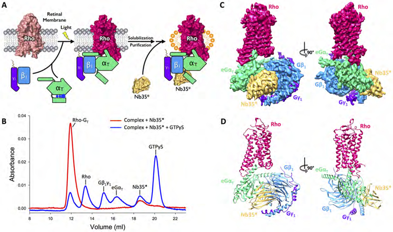
Figure 1. Purification and structural determination of the rhodopsin (Rho)-transducin (GT) complex.

(A) Schematic illustration of the purification of the Rho-GT complex. The complex is formed on retinal membranes through light activation of Rho, solubilized with detergent, and purified through chromatography steps. The engineered nanobody (Nb35*) is then added to the Rho-GT complex in excess. (B) Size exclusion chromatography (SEC) profiles of the purified Rho-GT-Nb35* complex (red) and its dissociation upon the addition of GTPγS (blue). (C) Orthogonal views of the cryo-EM density map colored by subunit (Rho in strawberry, eGαT in lime, Gβ1 in blue, Gγ1 in purple, Nb35* in gold). (D) Structure of the Rho-GT-Nb35* complex in the same views and color scheme.
See also Figures S1–S3 and Table S1.
