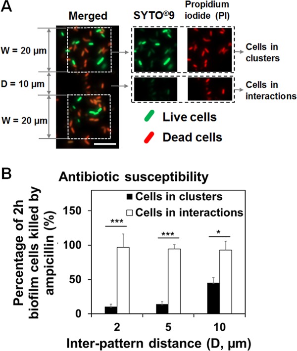
FIG 3.

Cell-cell interactions affected the Amp susceptibility of attached cells. (A) Representative fluorescence images of 2-h patterned biofilms treated with 200-μg/ml Amp and labeled with live/dead staining (bar, 10 μm). The antibiotic treatment was conducted in 0.85% NaCl solution. (B) Percentage of 2-h patterned biofilm cells (W = 20 μm and D = 2, 5, or 10 μm) killed by 200-μg/ml Amp (*, P < 0.05; **, P < 0.005; and ***, P < 0.0005). E. coli RP437 biofilms were formed on gold-coated glass surfaces in LB medium, and each condition was tested with three biological replicates.
