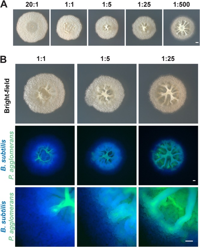
FIG 3.

Colony morphology and localization of B. subtilis and P. agglomerans in coculture plated at various initial cell ratios. (A) Colonies of B. subtilis and P. agglomerans in coculture imaged from the top and side at increasing initial ratios of P. agglomerans after growth on MSgg for 48 h. Bar, 1 mm. (B) B. subtilis (false colored blue) and P. agglomerans (false colored green) in coculture at increasing initial ratios of P. agglomerans grown on MSgg after 48 h of growth. Colocalization of two species is indicated in cyan. Coculture images of phase contrast (top) at ×1 magnification, fluorescence (middle) at ×1 magnification, and fluorescence (bottom) at ×8 magnification. Bars, 0.5 mm.
