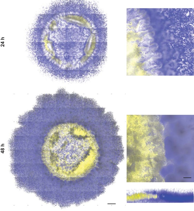
FIG 5.
B. subtilis and P. agglomerans are spatially organized within the coculture biofilm. Coculture colonies grown on glass coverslips embedded in MSgg agar and imaged at 24 h and 48 h using confocal laser scanning microscopy. B. subtilis PspacC-mTurq (blue) and P. agglomerans PspacC-Ypet (yellow) are shown in coculture colony tile scans (left) and an inset of one tile (right) with a representative three-dimensional (3D) reconstructed biofilm shown at 48 h. Bars, 500 μm and 100 μm for tile scans and individual tiles, respectively.

