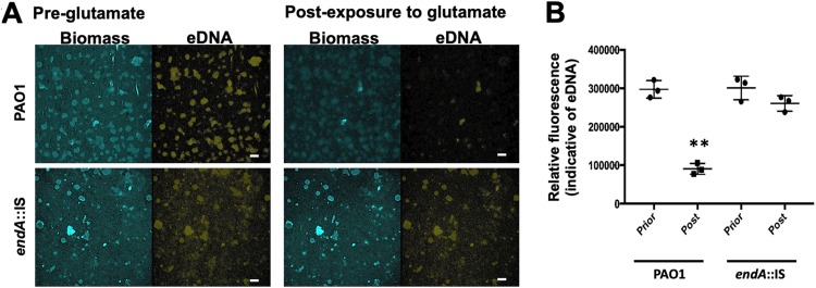
FIG 4.
EndA contributes to eDNA degradation in biofilm upon induction of dispersion. (A) Representative confocal images of 5-day-old biofilms by PAO1 and endA::IS strains prior to and after addition of the dispersion cue glutamate. eDNA was visualized using propidium iodide. Bars, 100 μm. (B) Quantitative analysis of the relative fluorescence associated with eDNA. Experiments were performed in triplicate, with each biological replicate consisting of 4 technical replicates. **, P < 0.05 relative to biofilms prior to exposure to glutamate (pre-glutamate).

