Fig. 4.
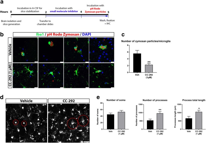
BTK inhibition decreases microglial phagocytosis and alters microglial morphology ex vivo. a Schematic diagram of the ex vivo microglial phagocytosis assay using acute rat brain slices. b Representative confocal images of acute rat brain slices incubated with 1 μM of CC-292 for 2 h followed by incubation with pHrodo® zymosan particles for 1 h. Uptake of zymosan particles by microglia was visualized by Iba1 immunostaining. Scale bar = 25 μm. c Number of zymosan particles phagocytosed per microglia was significantly reduced following CC-292 treatment. d Representative Axioscan images of Iba1-positive microglia in acute rat brain slices incubated with CC-292. Scale bar = 50 μm. e Measurements of microglia number and morphology showed significantly increased number of microglial processes and increased process length in CC-292-treated brain slices. Data presented as mean ± s.e.m., n = 5–6 slices per condition, unpaired t-test, *p < 0.05, **p < 0.01 versus vehicle control

