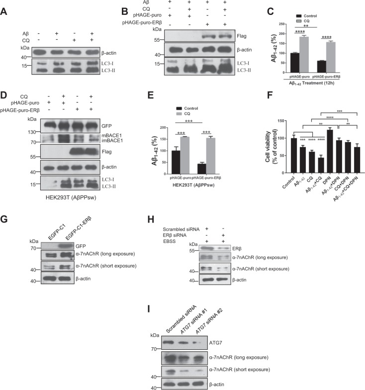
Fig. 6. Enhanced autophagy promotes extracellular Aβ1–42 degradation in SH-SY5Y cells.
a Cells were treated with Aβ (1 μM), CQ (10 μM), and Aβ plus CQ for 12 h. Cell lysates were analyzed by immunoblotting for LC3-II and β-actin protein expression. b After cells were transiently transfected with pHAGE-puro and pHAGE-puro-ERβ for 24 h, cells were treated with CQ 10 (μM) for 12 h. Next, cells were treated with Aβ1–42 Aβ fibrils for 12 h. LC3-II level was tested by western blot. c Cells were treated as described in b, the Aβ1–42 concentration was measured by an ELISA assay. d Cells were transfected with ERβ and vector plasmids under CQ treatment or not in the HEK293T (AβPPsw) model. Cell lysates were analyzed by immunoblotting for APP, BACE1, Flag, LC3-II, and β-actin protein expression. e Cells were treated as described in d, the Aβ1–42 concentration was measured by an ELISA assay. f Cells were treated with DPN (10 nM) or Aβ1–42 (5 μM) for 12 h and then added CQ (10 μM) for another 12 h. The MTT assay was used to test cell viability. g Cells were treated as described in (Fig. 1d), then the α7nAChR level was tested by western blot. h Cells were treated as described in (Fig. 1b), then the α7nAChR level was tested by western blot. i Cells were treated as described in (Fig. S2A), then the α7nAChR level was tested by western blot. Data shown are mean ± S.D. of three independent experiments. (*P < 0.05; **P < 0.01; ***P < 0.001; ****P < 0.0001)

