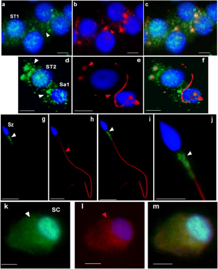
Fig. 5.
Immunocytochemical detection of CCDC103 (green) and of axoneme-specific acetylated α-tubulin (red), with merged images, in cells of the seminiferous tubules from controls (i.e., samples obtained from men with conserved spermatogenesis that are under infertility treatments). Staining was observed in the cytoplasm of primary spermatocytes (ST1: a–c), secondary spermatocytes (ST2: d–f), and round spermatids (Sa: d–f), and in the midpiece of sperm (Sz: g–j). In culture Sertoli cells (SC: k–m), staining was observed in the cytoplasm and in the perinuclear region. Nuclei stained with DAPI (blue). White arrowheads-CCDC103 staining, red arrowheads-tubulin staining. Scale bars, 5 μm

