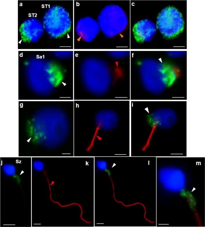
Fig. 6.
Immunocytochemical detection of CCDC103 (green) and of axoneme-specific acetylated α-tubulin (red), with merged images, in cells of the seminiferous tubules from patient 1. Staining was observed in the cytoplasm and perinuclear region of primary spermatocytes (ST1: a–c) and secondary spermatocytes (ST2: a–c), at the basal pole of the nucleus preceding axoneme extrusion in early round spermatids (Sa1: d–f) and late round spermatids (Sa2: g–i), and (granular appearance) in the midpiece of sperm (Sz: j–m). Nuclei stained with DAPI (blue). White arrowheads, CCDC103 staining; red arrowheads, tubulin staining. Scale bars: a–c, 4 μm; d–f, 2 μm; h, i, 4 μm; g, 2 μm; j–m, 2 μm

