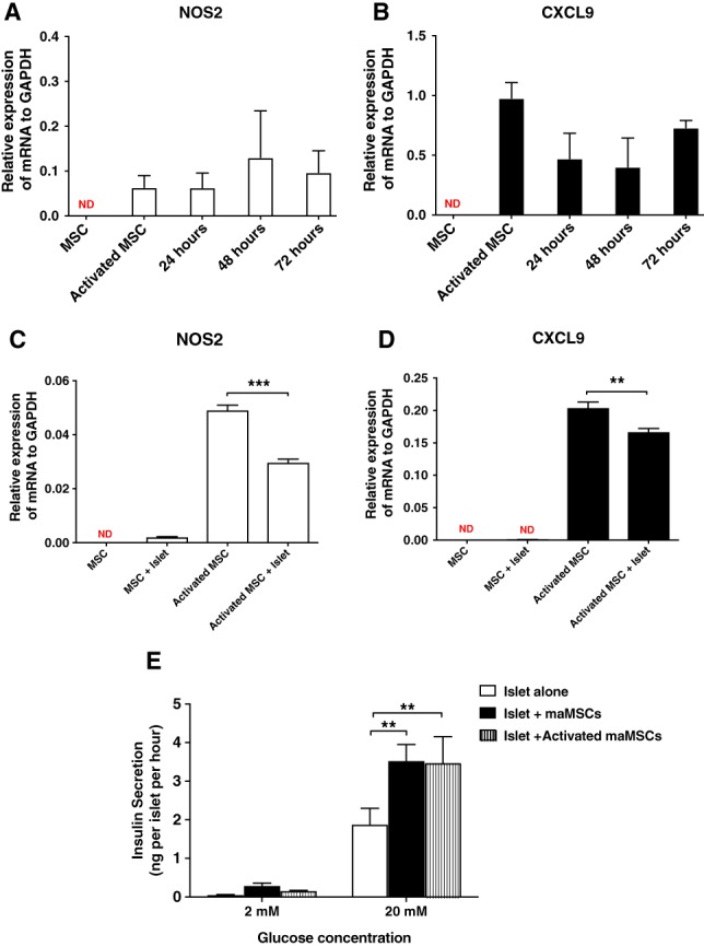
Figure 4.

Activation of maMSCs. (A, B): Cytokine‐induced activation of maMSCs. maMSCs were cultured in the absence or presence of interferon‐γ (20 ng/ml) and tumor necrosis factor‐α (20 ng/ml) for 8 hours and NOS2 (A) and CXCL9 (B) mRNAs were measured 24, 48, and 72 hours after the removal of cytokines. Data are expressed as mean + SEM, n = 3 in one experiment representative of three separate experiments. **, p < .001; ND, not detectable. (C, D): Effects of islet coculture on maMSC activation. Quiescent or cytokine‐activated maMSCs were cocultured with mouse islets for 72 hours, followed by the measurement of NOS2 (C) and CXCL9 (D) mRNAs in the maMSC populations. Data are expressed as mean + SD; n = 3 observations in one experiment representative of three separate experiments. **, p < .01. (E): Effects of maMSC activation on glucose‐stimulated insulin secretion from mouse islets. Insulin secretion at substimulatory (2 mM) and stimulatory glucose concentration (20 mM) from islets alone (white bar), islets cocultured with maMSCs (black bar), and islets cocultured with activated maMSCs (hatched bar). Data are presented as mean + SEM; n = 10 observations. ***, p < .001; **, p < .01; *, p < .05. Abbreviations: GAPDH, glyceraldehyde‐3‐phosphate dehydrogenase; maMSCs, mouse adipose mesenchymal stromal cells; MSCs, mesenchymal stromal cells.
