Figure 3.

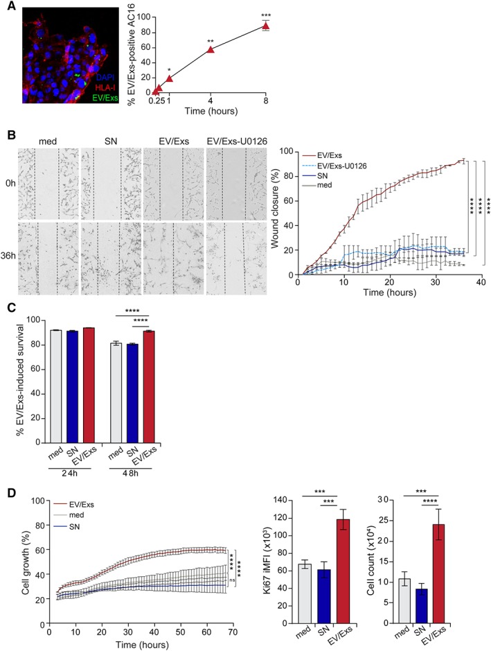
Internalization of extracellular vesicles/exosomes (EV/Exs) by AC16 cardiomyocytes promotes their migration, survival, and proliferation. (A): Uptake of carboxyfluorescein N‐succinimidyl ester‐labeled EV/Exs by AC16 cardiomyocytes as assessed by fluorescent microscopy (left panel, ×200 magnification) and flow cytometry at different time points as indicated (right panel). Results are mean values ± SD from three independent experiments. (B): EV/Exs and non‐EV/Exs‐free medium (SN) or medium alone (med) promote AC16 cardiomyocytes migration, which is blocked in the presence of U0126. Representative images (×10 magnification) of four independent scratch wound assays at 0 and 36 hours (left panel), and the %wound closure as monitored over time and calculated by ImageJ software. Results are presented as mean values ± SD from two independent experiments. (C): Survival of 12‐hours starved AC16 cardiomyocytes in fetal bovine serum (FBS)‐free medium in the absence (med) or the presence of EV/Exs or EV/Exs‐free medium (SN) as determined by cell viability dye at 24 and 48 hours. Results are presented as mean values ± SD from three independent experiments. (D): AC16 cardiomyocytes proliferation in FBS‐free medium in the absence (med) or presence of EV/Exs or EV/Exs‐free medium (SN) as evaluated by monitoring cell growth over 70 hours in Incucyte (left panel), and staining for Ki67 proliferation marker (middle panel) and cell counting using flow cytometry (right panel) at 24 hours. Results are presented as mean value ± SD of %cell growth, Ki67 integrated mean fluorescence intensity, and number of cells, respectively, from three independent experiments. *, p < .05; **, p < .01; ***, p < .001; ****, p < .0001 compared with med or SN.
