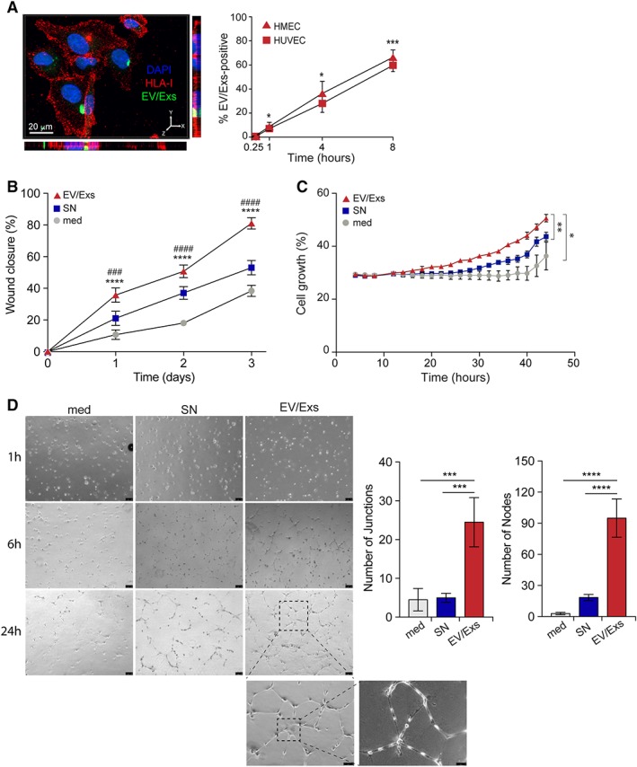
Figure 4.

Internalization of extracellular vesicles/exosomes (EV/Exs) by human microvascular endothelial cells (HMEC) and human umbilical vein endothelial cells (HUVEC) promote migration, growth, and tube formation. (A): Representative Z‐stack projections of immunofluorescence image by Imaris program for carboxyfluorescein N‐succinimidyl ester (CFSE)‐labeled EV/Exs uptake by HUVEC. Orthogonal views are shown with Y–Z and X–Z orientations (left panel). In the right panel, the uptake of CFSE‐labeled EV/Exs by HUVEC and HMEC as assessed by flow cytometry at different time points as indicated. (B): EV/Exs and non‐EV/Exs‐free medium (SN) or medium alone (med) promote HUVEC migration. The %wound closure as monitored on over time as indicated and calculated by ImageJ software. Similar data were obtained with HMEC. (C): Serum‐starved HMEC growth in the absence (med) or presence of EV/Exs or EV/Exs‐free medium (SN) monitored over 48 hours in Incucyte and assessed by integrated software. Similar data were obtained with HUVEC. (D): Organization of HUVEC in tube structures in the absence (med) or presence of EV/Exs or EV/Exs‐free medium (SN) over 24 hours. Representative images of three independent experiments (left panel, ×40, ×100, and ×200 magnification) and histograms presenting number of nodes and junctions as determined by angiogenesis analyzer for ImageJ software (right panel). All results are presented as mean values ± SD from at least two independent experiments. *, p < .05; **, p < .01; ***, p < .001; ****, p < .0001 compared with medium; ###, p < .001; ####, p < .0001 compared with SN.
