Figure 3.

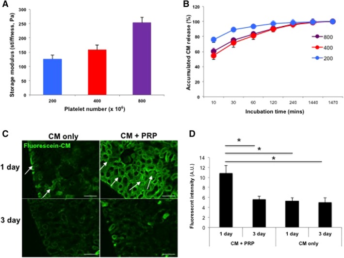
Controlled release of CM from PRP gel in vitro and in vivo. (A): Mechanical stiffness of PRP gels increased with increased concentrations of platelets (n = 3 per sample). (B): Sustained release of CM from all PRP gels was detected over 24 hours, and stiffer gels (400 and 800 × 106 platelets) released CM more slowly. Based on these results, PRP with 400 × 106 platelets were chosen for all further experiments. (C): Fluorescently labeled CM encapsulated in the PRP gel was localized to cortex region (arrows) at 1 day after ischemia/reperfusion, whereas CM without PRP quickly disappeared. In both groups, intensity of green fluorescence gradually decreased up to 3 days. (D): Semiquantitative analysis for fluorescent intensity showed more effective delivery of CM with PRP gel than CM‐only injection at 1 day postinjection (*p < .05). Data presented as mean ± SD (n = 2 animals, four kidneys per experimental group; ×200 magnification, Bar = 100 μm). Abbreviations: A.U., arbitrary units; CM, conditioned medium; PRC, primary renal cell.
