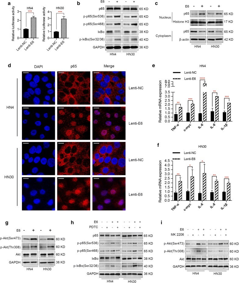
Fig. 3.
E6 oncogene activates NF-κB and Akt pathways in HNSCC. a NF-κB luciferase reporter assay demonstrated an increase of NF-κB activities in E6-expressing HNSCC cells, suggesting the activation of NF-κB pathway by E6 oncogene. b Western blot results illustrated that E6 oncogene activated NF-κB pathway and regulated signaling-related proteins expression. c Western blot analysis revealed that p65 was localized mostly in the cytoplasm in E6 negative HNSCC cells, while p65 was translocated from the cytoplasm to the nucleus in E6-expressing cells. d Confocal microscopy analysis confirmed the nuclear accumulation of p65 in E6-expressing HNSCC cells. e, f mRNA levels of several key pro-inflammatory NF-κB-dependent cytokines and genes TNF-α, IL-1β, IL-6, IL-8 and c-myc were elevated in E6 positive HNSCC cells. g E6 oncogene activated Akt signaling pathway in HNSCC, demonstrated by promoting the phosphorylation of Akt protein. h, i Specific inhibitors of NF-κB (PDTC, Beyotime) and Akt pathways (MK-2206, Selleck) were utilized to demonstrate the effect of E6 oncogene on HNSCC was really dependent on these two pathways. *P < 0.05. **P < 0.01. ***P < 0.001. ****P < 0.0001 (scale bar: 10 μm)

