Fig. 4.
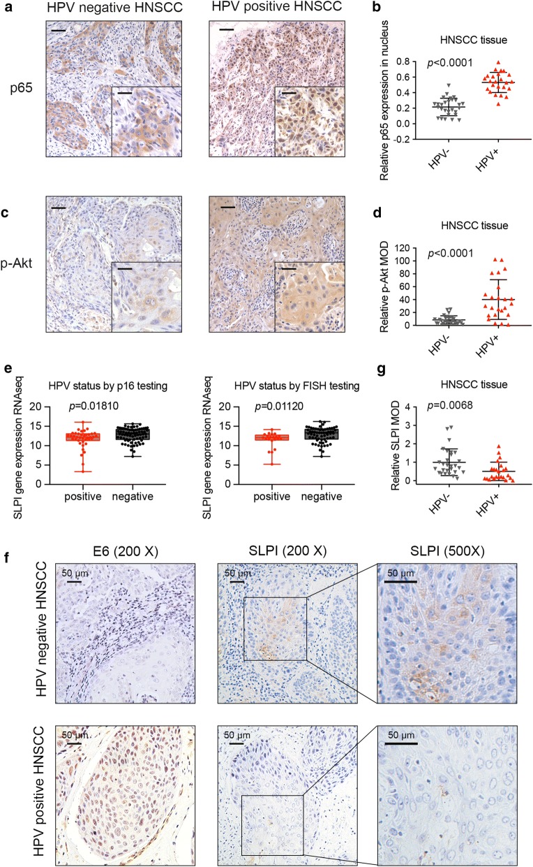
Relative activation of NF-κB and Akt pathways and SLPI downregulation in HPV positive HNSCC. a, b The proportion of p65 expressed in the cell nucleus in HPV positive tissues was significantly higher than that in HPV negative tissues. c, d The expression level of p-Akt in HPV positive HNSCC samples was much higher when compared to that in HNSCC samples without HPV infection (scale bar: main = 50 μm; insert = 15 μm). e mRNA expression level of SLPI was significantly decreased in HPV positive HNSCC samples (diagnosis both by FISH testing and p16 testing) according to the analysis results of mRNA data from the Cancer Genome Atlas (TCGA, http://xenabrowser.net). f Immunohistochemistry assay showed that HPV positive HNSCC tissues displayed a widespread expression of E6 oncogene, both in nuclear and cytoplasm while HPV negative tissues presented no E6 expression. Meanwhile, the staining intensity of SLPI was obviously lower in HPV positive HNSCC when compared to HPV negative HNSCC. g Statistical analysis of immunohistochemistry conducted on 24 HPV positive HNSCC tissues and 28 HPV negative tissues illustrated that SLPI protein level in HPV positive HNSCC was statistically lower than that in HPV negative ones

