Fig. 5.
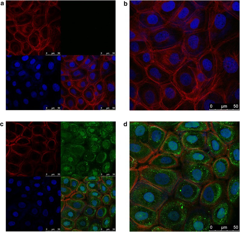
Exogenous SLPI could get internalized into HNSCC cells. a, b The images of HN4 cells incubated without exogenous SLPI protein. c, d The images of HN4 cells incubated with 40 μg/mL exogenous SLPI protein for 1 h. Cell nucleus was stained with DAPI (blue). Cytoskeleton was stained with phalloidine (red). SLPI was stained with FITC secondary antibody (green) (scale bar: 50 μm)

