Fig. 2.
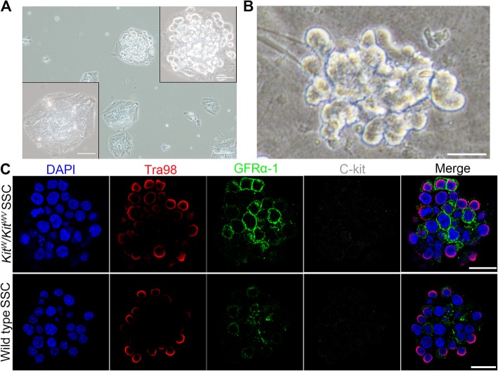
The isolating strategy and characterization of Kitw/Kitwv SSCs. a All tested testes were digested separately. With the lack of germ cells, most digested testes samples were tiled in the bottom of the dish like the lower left panel after being digested into small fragments and cultured for 18 h. A small number of testes have rare tubules containing germ cells presented as the upper right panel. Testes samples containing germ cell stacks were selected for SSC enrichment. Scale bar = 100 μm in original size panels and 50 μm in magnified panels. b Morphology of isolated Kitw/Kitwv SSC clump. Scale bar = 20 μm. c Identification of SSCs. Kitw/Kitwv mutant SSC clumps (DAPI, for cell nuclei) were positive for germ cell marker Tra98 (red) and stem cell GFRα-1 (green), but negative for differentiated germ cell C-kit (gray), which were consistent with the wild type control SSCs shown in the bottom panel of c, indicating that they were undifferentiated spermatogonial stem cells. Scale bar = 20 μm

