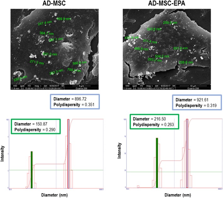
Fig. 3.
Upper panels: Scanning electron microscopy of adipose tissue (AD)-derived mesenchymal stromal cells (MSCs). Note the presence of extracellular vesicles on AD-MSC surfaces. Lower panels: Representative graph of the intensity and hydrodynamic diameter of extracellular vesicle samples, analyzed using the dynamic light scattering technique. Graph shows two populations of extracellular vesicles obtained from nonpreconditioned (AD-MSC) and EPA-preconditioned MSCs (AD-MSC-EPA): one of lower intensity and medium size, characteristic of exosomes, and another with greater intensity and average size, characteristic of microvesicles. Green and blue lines denote the mean diameter

