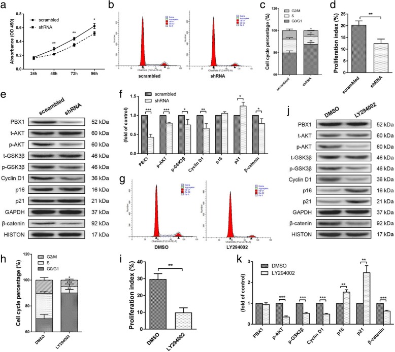
Fig. 3.
Knockdown of PBX1 in HF-MSCs suppressed proliferation and inhibited the AKT/GSK3β pathway. a Cell proliferation curves for HF-MSCsscrambled and HF-MSCsshRNA. b, c Percentages of cells in the G1, S, and G2 phases of the cell cycle and PIs (d) for the HF-MSCsscrambled and HF-MSCsshRNA. e, f Western blot analysis of the levels of phospho-AKT, phospho-GSK3β, cyclin D1, p16, p21, and β-catenin proteins in HF-MSCsscrambled and HF-MSCsshRNA. g, h Percentages of cells in the G1, S, and G2 phases of the cell cycle and PIs (i) for HF-MSCsPBX1 cultured with DMSO and LY294002. j, k Western blot analysis of the levels of phospho-AKT, phospho-GSK3β, cyclin D1, p16, p21, and β-catenin proteins in HF-MSCsPBX1 cultured with DMSO and LY294002. *P < 0.05; **P < 0.01; ***P < 0.001

