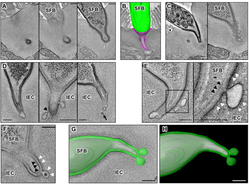
Fig. 1. Microbial Adhesion-Triggered Endocytosis (MATE) is induced following attachment of commensal segmented filamentous bacteria (SFB) to intestinal epithelial cells (IECs).
(A) Consecutive sections of an electron tomogram of an SFB-IEC synapse showing that double membrane phagosome-like vesicles (left panel) represent invaginations of the IEC plasma membrane. (B) A 3D reconstruction of an SFB-IEC holdfast, demonstrating separation of the IEC plasma membrane (PM) in purple and the SFB PM in green/gray. SFB do not penetrate the IEC PM. (C) Membrane vesicles at the tip of SFB holdfasts. (D) Holdfast vesicles form necks (arrowhead) and bud off (arrow) of the IEC PM into the IEC cytosol. (E, F) SFB PM (black arrowheads) remains uninterrupted and holdfast vesicles form exclusively from the host IEC PM (white arrowheads) and contain electron dense cargo (F). (G, H) Reconstruction of an SFB holdfast. IEC PM in green, SFB PM in gray. All scale bars are 200 nm.

