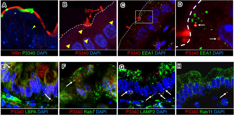
Fig 3. SFB antigens are shuttled through the IEC endosomal-lysosomal vesicular network.
(A, B) Immunofluorescence for P3340 on intestinal sections from terminal ileum showing SFB P3340 inside IECs (yellow arrowheads). (C, D) P3340 is present in EEA1+ early endosomes. White dashed line outlines the apical IEC surface. (E-H) Co-localization of intracellular SFB protein P3400 (red) with endosomal and lysosomal markers (green). P3340 co-localizes with late endosomes (LBPA, Rab7) (E, F), as well as basolateral lysosomes (LAMP2) (G). (H) Some P3340 also co-localizes with recycling endosomes (Rab11).

