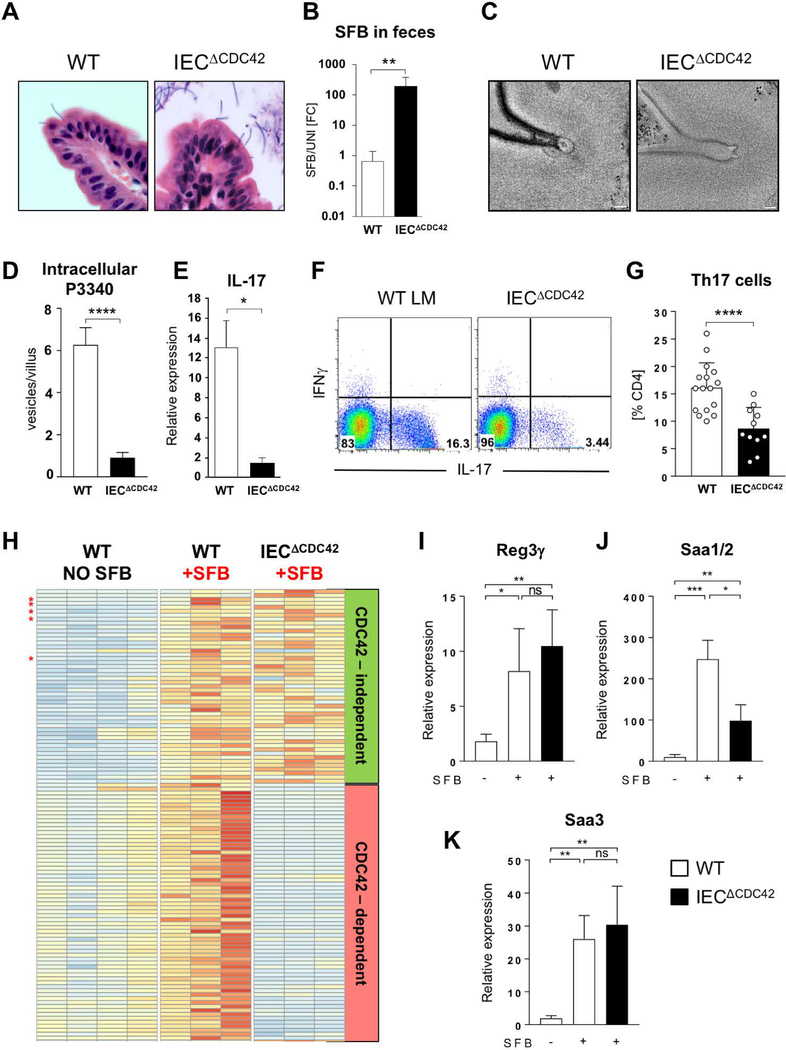
Fig 7. Epithelial CDC42 is required for MATE, SFB antigen acquisition and Th17 cell induction by SFB.
(A) H&E staining of sections from terminal ileum of WT and IECΔCDC42 mice. SFB attachment on the surface of the villi is evident in both groups. (B) SFB levels in feces from WT and IECΔCDC42 mice. (C) MATE vesicles in SFB-IEC synapses in terminal ileum of WT and IECΔCDC42 mice. (D) Decrease in acquisition of P3340 by IEC in terminal ileum of IECΔCDC42 mice. (E) Decrease in IL-17 mRNA in terminal ileum of SFB-colonized IECΔCDC42 mice. (F, G) Decrease in small intestinal lamina propria (LP) Th17 cells in IECΔCDC42 mice. Plots in (F) gated on TCRβ+CD4+ lymphocytes. (G) Combined data from several independent experiments. (H) Relative expression levels of SFB-induced genes in IECs from WT and IECΔCDC42 mice. Only genes induced by SFB are depicted. Red stars indicate Saa1, Saa2, Saa3, Nos2, and Reg3g. Complete list of gene names is included in Fig. S9. (I-K) RT-PCR for SFB-controlled genes on RNA isolated from IECs from WT and IECΔCDC42 mice as described in Materials and Methods. Statistics, unpaired t test. * p < 0.05, ** p < 0.01, *** p < 0.005, **** p < 0.001. Scale bars in (C) are 100 nm.

