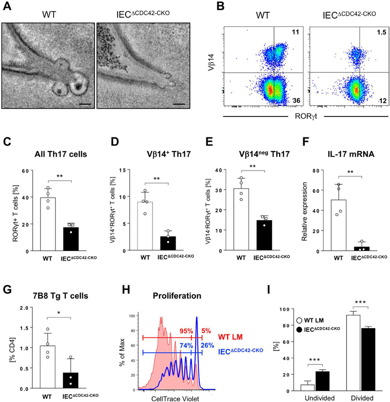
Fig. 8. Epithelial CDC42 is required for activation of SFB-specific CD4 T cells and induction of SFB-specific Th17 cells.
WT and IECΔCDC42-CKO mice were treated with tamoxifen prior to SFB colonization and transfer of naïve P3340-specific 7B8 Tg CD4 T cells (for details see Materials and Methods). (A) MATE vesicles in SFB-IEC synapses in terminal ileum of tamoxifen-treated WT and IECΔCDC42-CKO mice. (B-E) Decrease in endogenous SI LP Th17 cells in tamoxifen-treated IECΔCDC42-CKO mice six days after SFB colonization. (B) Representative FACS plots of LP lymphocytes gated on TCRβ+CD4+ cells. (C-E) Statistic based on the gating in (B). (F) Decrease in IL-17 mRNA in terminal ileum of tamoxifen-treated IECΔCDC42-CKO mice. (G) Decreased expansion of adoptively transferred P3340-specific 7B8 Tg CD4 T cells in mesenteric lymph nodes (MLN) of tamoxifen-treated IECΔCDC42-CKO mice four days after transfer. (H, I) Proliferation of 7B8 Tg CD4 T cells in MLN of tamoxifen-treated WT littermate (WT LM) or IECΔCDC42-CKO recipient mice on Day 4 after adoptive transfer. One of three independent experiments with similar results. Error bars, standard deviation. Statistics, unpaired two-tailed t test. * p < 0.05, ** p < 0.01, *** p < 0.005, **** p < 0.001. Scale bars in (A) are 100 nm.

