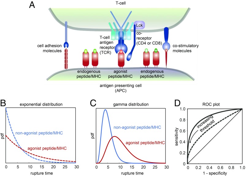
Fig. 1.
(A) Scheme outlining T cell antigen recognition in the context of the immunological synapse, the area of contact between a T cell and an APC. T cells manage to detect the presence of even a single agonist pMHC complex among highly abundant nonstimulatory but structurally similar endogenous pMHC complexes. Forces acting on TCR–pMHC bonds may help in discriminating between activating and nonactivating pMHC. Previously proposed catch bonds between TCR and activating ligands were not observed in the recent study by Limozin et al. (13), highlighting the importance of slip bonds for effective and sensitive detection of rare agonist ligands. Note the presence of coreceptors (tethered to Lck, an intracellular kinase interacting with the TCR upon ligand engagement), adhesion molecules, and costimulatory molecules within the immunological synapses with possible implications for TCR–pMHC engagement. (B–D) Classification according to rupture times. (B) Comparison of 2 exponential rupture time distributions with a mean of 5 (blue) and 10 (red). (C) Comparison of 2 gamma-distributed rupture time distributions with the same mean values as in B. (D) ROC plot for the exponential (dashed line) and the gamma-distributed (solid line) rupture time distribution under variation of the discrimination threshold. With increasing threshold, specificity increases at the expense of a decreasing sensitivity.

