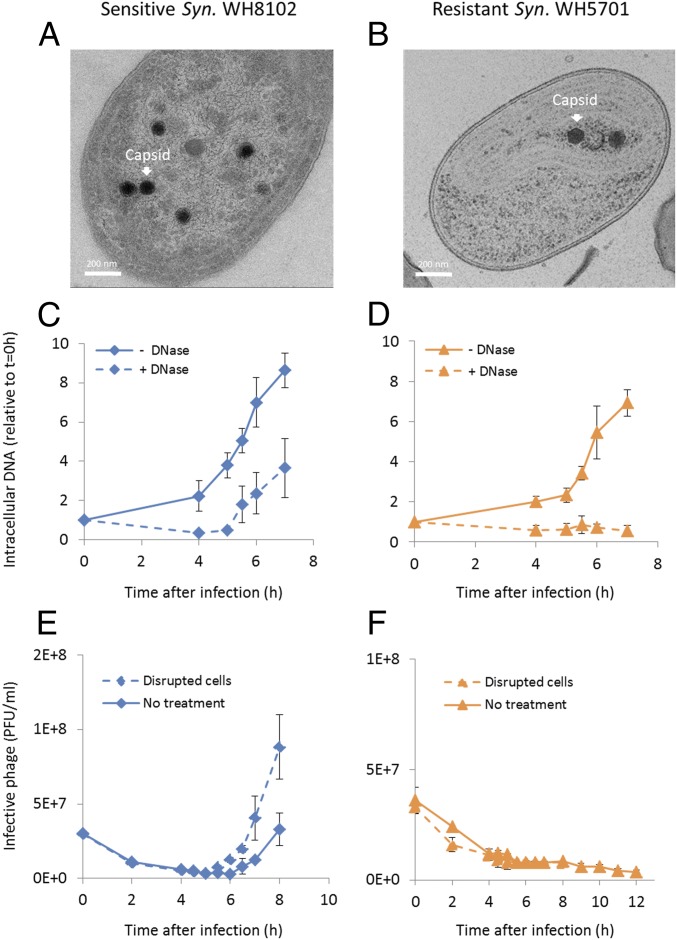
Fig. 5.
Formation of Syn9 phage particles in resistant Synechococcus WH5701. Capsid assembly is observed from TEM images of thin cell sections (A and B), packaging of DNA into capsids determined from DNase protection (C and D), and intracellular production of infective phage determined from plaque-forming units (E and F) during Syn9 infection of sensitive Synechococcus (Syn.) WH8102 (A, C, and E) and resistant Synechococcus WH5701 (B, D, and F) strains. TEM images are representative of 2 independent experiments (n = 18 sections for Synechococcus WH8102 and n = 28 sections for Synechococcus WH5701). Size distribution of phage particles is shown in SI Appendix, Fig. S5. (C and D) “− DNase” treatment indicates total intracellular DNA, while “+ DNase” treatment indicates intracellular DNA that is protected from DNase digestion after cell disruption (n = 3). Phage DNA levels were determined by qPCR for the g20 portal gene. (E and F) “Disrupted cells” treatment shows the number of infective phages present intracellularly and in the extracellular medium, while “No treatment” shows the number of infective phages in the extracellular medium (n = 3). PFU, plaque-forming units.

