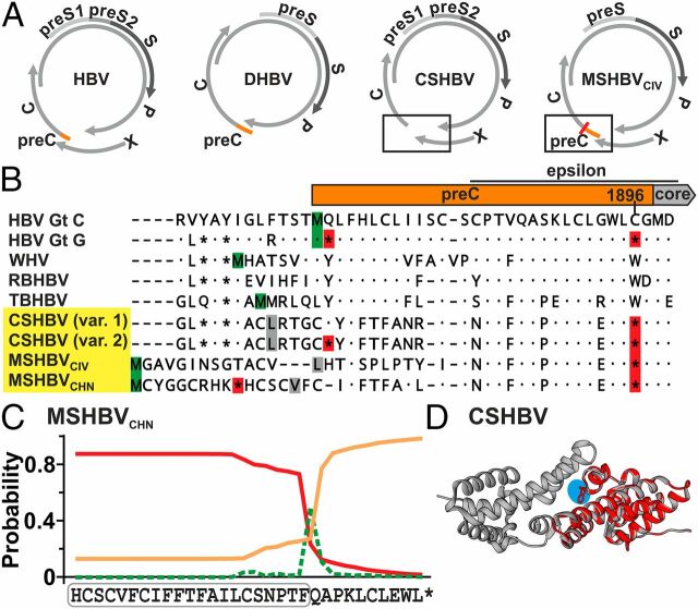
Fig. 3.
Shrew HBV HBeAg. (A) Genome structures. (B) Translated preC and N-terminal core domains. Red, stop codons; green, methionine; gray, alternative start codons. Var. 1/2, HTS minority variants (25–30% occurrence). (C) Signal peptide prediction (red line) of MSHBVCHN. Green, cleavage site; orange line, no signal peptide; boxed, predicted signal sequence. (D) CSHBV HBeAg monomer (cyan, reverted G1896A) modeled on the HBV HBeAg dimer (43). Gt, genotype; RBHBV, roundleaf bat HBV.

