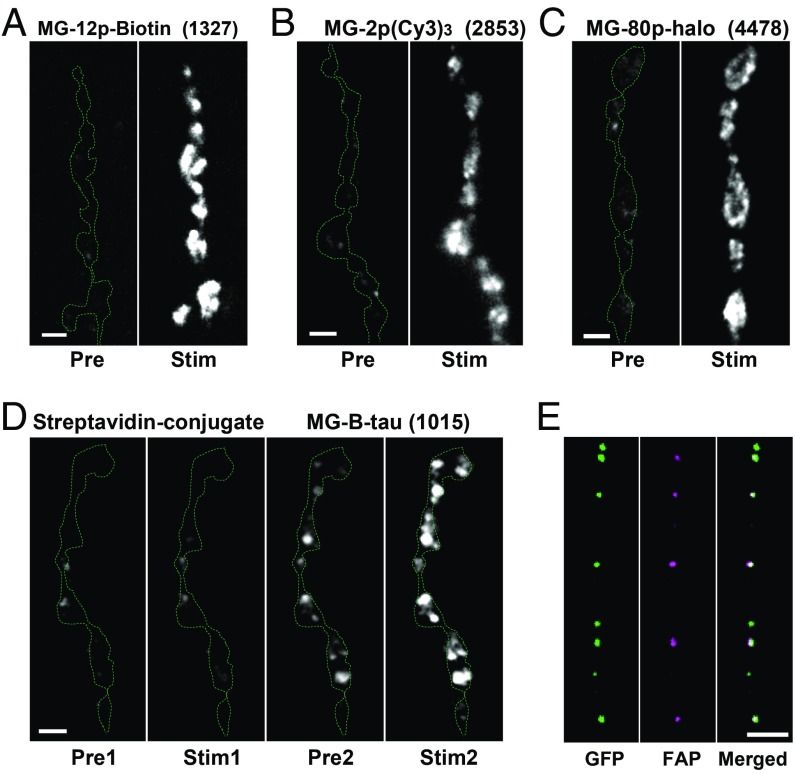
Fig. 3.
FAP labeling is mediated by fusion pores. Representative images of Dilp2-FAP expressing boutons before (Pre) and after 70 Hz stimulation for 1 min (Stim) in the presence of MG-12p-biotin (A), MG-(Cy3)3 (B), and MG-p80 (C). Boutons outlined with the dashed line. Numbers in parentheses are MW in daltons of the MG derivative. (D) Representative mages of Dilp2-FAP expressing boutons in the presence of MG-12p-biotin conjugated with streptavid before (Pre1) and after 70 Hz stimulation for 1 min (Stim1) and after replacing the conjugate with MG-B-tau (Pre2) and after 70 Hz stimulation for 1 min (Stim2). (E) Pseudocolor images of DCVs in the axon coexpressing Dilp2-FAP (magenta) and Dilp2-GFP (green) fixed in paraformaldehyde 5 min after 70 Hz stimulation for 60 s in the presence of MG-2p (Scale bars, 2 µm.)

