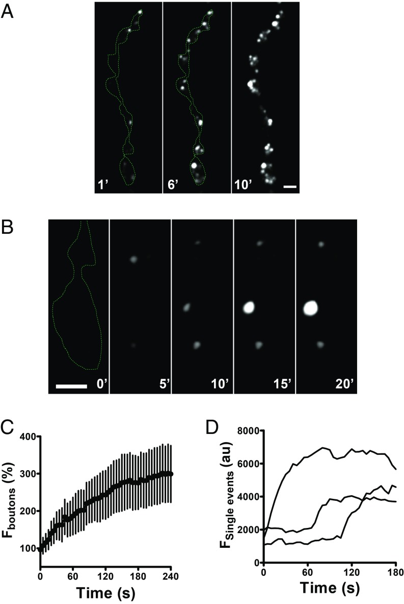
Fig. 4.
Spontaneous fusion pore openings in Dilp2-FAP expressing boutons in the presence of MG-2p. (A) Representative images of Dilp2-FAP expressing boutons in the absence of Ca2+ after application of MG-2p. Numbers on the images indicate minutes. (B) Consecutive images of Dilp2-FAP expressing boutons in the absence of Ca2+after application of MG-2p show that individual puncta vary in whether they grow brighter with time. Numbers on the images indicate minutes (Scale bars, 2 µm.) (C) Time course of bouton fluorescence increase in the absence of Ca2+ after application of MG-2p. Data are from 5 animals (12 boutons). (D) Representative time courses of individual spontaneous fusion pore openings in the absence of Ca2+ after application of MG-2p.

