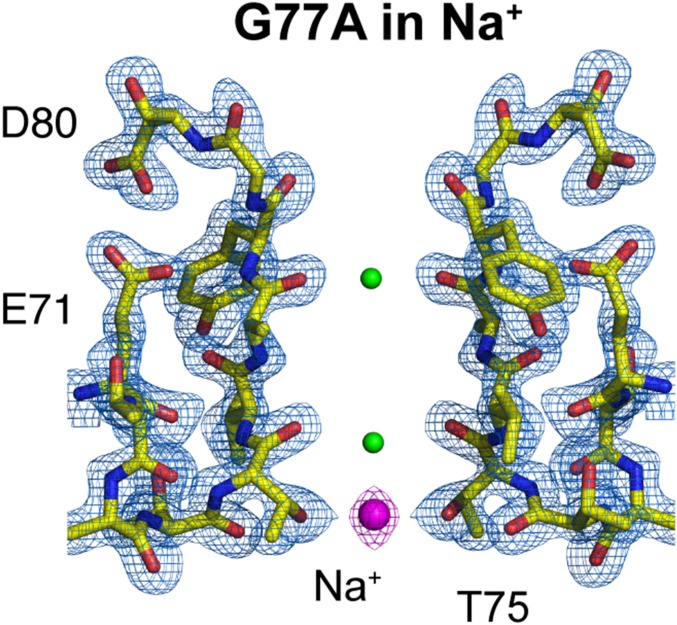
Fig. 4.
Crystal structure of the G77A mutant in 300 mM sodium chloride. The G77A structure solved in the presence of 300 mM NaCl at 2.05-Å resolution (PDB is 6PA0). A 2Fo-Fc electron-density map (light blue, contoured at 2.6 σ) validates the SF structural model colored in yellow. One Na+ ion is bound to the S4 site by the carboxyl group of Thr75 (presented as magenta spheres). Two small positive electron densities were modeled as water molecules (represented as small green spheres).

