Figure 1.
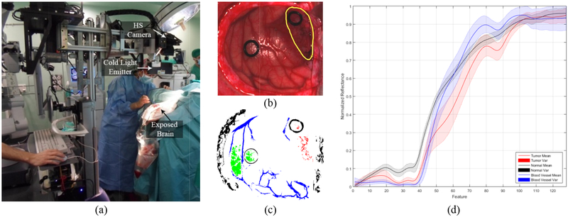
(a) Intraoperative HS acquisition system capturing an image during a surgical procedure. (b) Synthetic RGB etation of a HS cube from an in-vivo brain surfac unded in yello affected by GBM tumor (surro). (c) Golden wrepresen ith the semi-autmstandard wap obtained omatic labelling tool from the same HS cube. Normal, tumor, hypervascularizedand background classes are represented in green, red, blue and black color respectively. (d) Average and standard deviatiof the spectral signatures of the tumor (red), normal (black) and blood vessel/hypervascularized (blue) labelled pixels.

