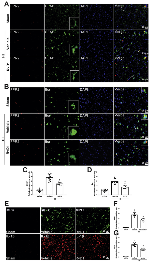
Fig. 4.
Localization of FPR2 and Iba1 (microglial marker) and GFAP (astrocytic marker) in the brain at 24 h post hypoxia-ischemia (HI). (A and B) Immunofluorescent staining showed that FPR2 was co-localized with Iba1 and GFAP. Scale bar=25 μm. (C and D) Quantitative analysis showed that more Iba1 and GFAP were expressed in HI+vehicle group, while RvD1 attenuated the increase. Representative pictures and schematic diagram showed there was more inflammation in vehicle group compared to sham, indicative by positive MPO and IL-1β (E-G), however, RvD1 treatment reduced the increase (E-G). ANOVA followed by Tukey test was used for analysis. Data were shown as mean ± SD (n=3 per group). * P < 0.05 vs. sham; # P < 0.05 vs. HI+vehicle. RvD1, resolvin D1.

