
Figure 1.
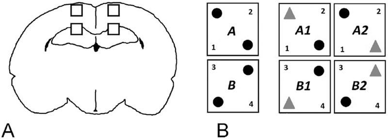
A: Histology Methods
Illustration of areas sampled for gliosis and microglia/macrophage phenotype in the M1 and CA1 regions of the cortex and hippocampus.
B: Novel Object Recognition Methods
Schematic representation of Novel Object Recognition testing paradigm: on the left, the two variations (A and B) on familiar object positioning (black spheres) during Familiarization. On the right, the two variations on novel object positioning (gray triangles in A1 and A2) or (B1 and B2) during Novel Object testing, as detailed in the text.